CIRS 603A头部模体,CIRS 603A头部MRI模体
型号:CIRS 603A 类别: 质控模体 品牌:美国CIRS pdf资料: CIRS 603A头部模体,CIRS 603A头部MRI模体.pdf
采购热线:0755-28896837 13632925349
在线咨询产品 
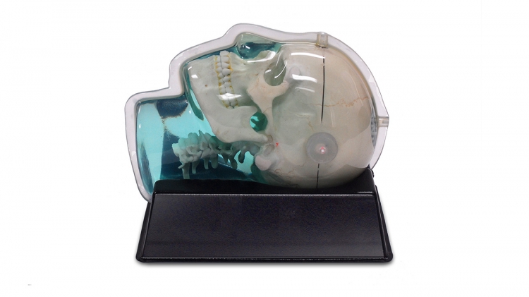
CIRS 603A头部模体由可用X射线,计算机断层扫描和磁共振成像技术成像的材料制成。它能很好地描述迄今为止所有测试的MRI序列,包括T1加权,T2加权,3D飞行时间,MPRAGE和CISS序列。
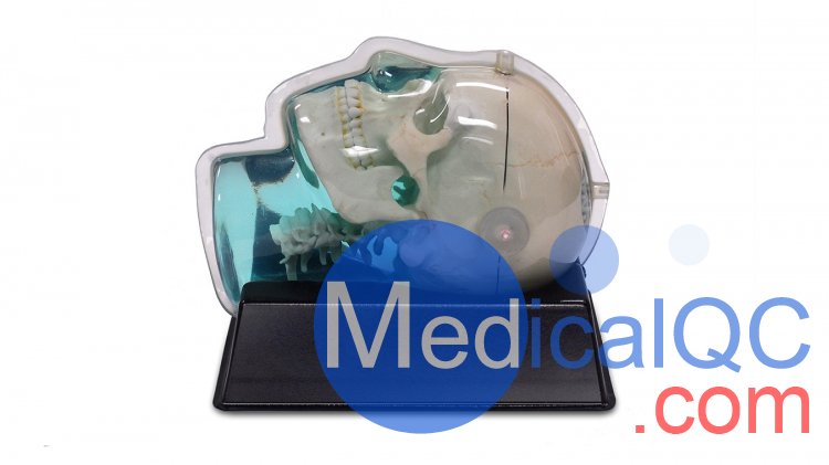
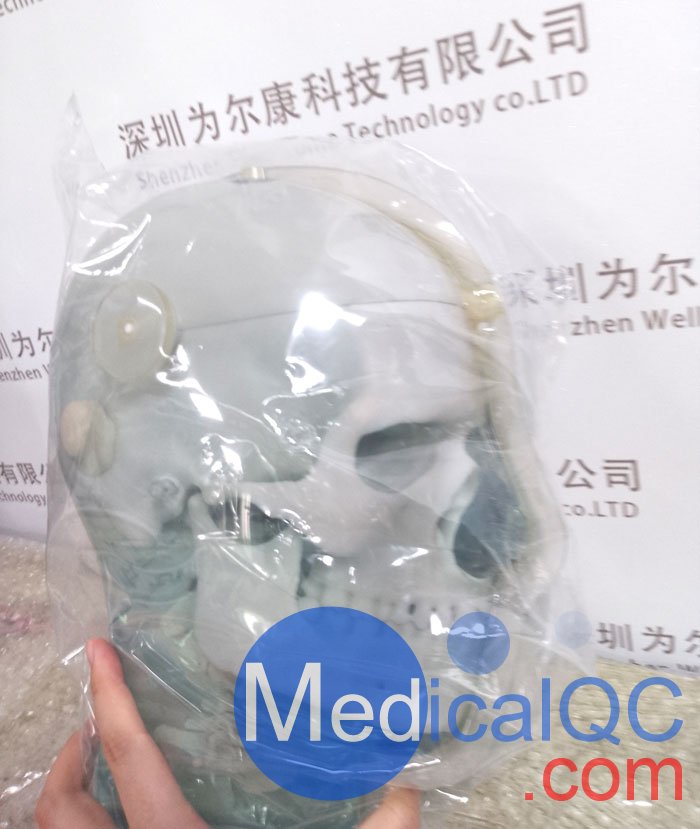
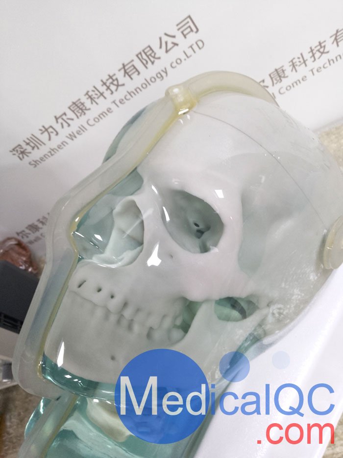
CIRS 603A头部模体的头骨是由塑料基组织替代品制造的,而间质和周围的软组织是由专有的水基聚合物制成的。整个模型装在一个清晰的真空成型塑料外壳中,以防止凝胶干燥。
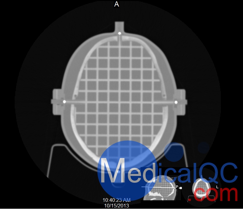
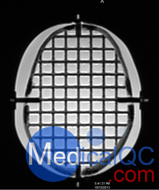
CIRS 603A头部模体的颅骨体的整个颅骨部分填充有间距为1.5cm的3mm直径杆的正交三维网格。五个扩展的轴杆在网格的参考原点相交。每个扩展轴的末端都配有CT和MR标记,可以准确对准激光以及图像配准。
CIRS 603A头部模体包括一个6.35毫米直径的丙烯酸棒,每个耳道有一个3毫米直径和17毫米长的空气空隙。
CIRS 603A头部模体特点和好处
T1,T2和3D TOF MRI采集的图像良好
CT扫描的图像很好
与立体定向框架一起使用的保护垫
图像可以导入立体定位程序
CT扫描可以用来评估MRI的准确性
包括的材料
3D拟人头骨幽灵
ABS真空成型摇篮
5立体框架垫
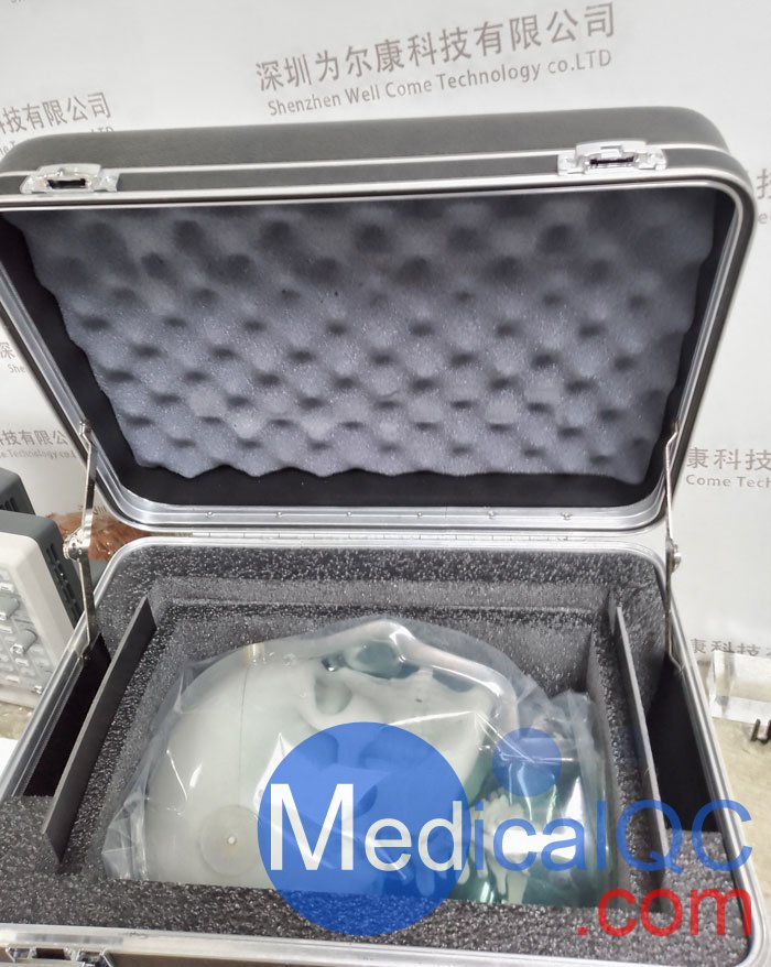
定制携带案例
用户指南
保修信息
4年保修
CIRS 603A was designed for assessment of MR image distortion in Stereotactic Radiosurgery Planning. It is also a useful tool for verifying image fusion and deformable image registration algorithms used in various treatment planning systems. The tissue equivalent, anthropomorphic design provides the closest conditions to a clinical imaging scenario. The phantom can be imaged using X-ray, Computed Tomography and Magnetic Resonance. It images well with all MRI sequences tested to date, including T1 weighted, T2 weighted, 3D Time of Flight, MPRAGE and CISS.
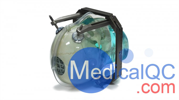
The skull is manufactured from a plastic-based bone substitute, and the interstitial and surrounding soft tissues are made from a proprietary signal generating water-based polymer. The entire phantom is encased in a clear plastic shell to protect gel from desiccation. The phantom is supplied with specially designed pads that allow fixation with any stereotactic frame or mounting for end-to-end testing. The phantom is also suitable for frameless SRS QA.
The entire inter-cranial portion of the skull volume is filled with an orthogonal 3D grid of 3mm diameter rods spaced 15 mm apart. Five extended axis-rods intersect at the reference origin of the grid. The end of each extended axis is fitted with CT/MR markers allowing for accurate positioning with lasers and co-registration of CT and MR image sets.
The phantom includes right and left air voids, 3 mm in diameter by 17 mm long to simulate each ear canal for evaluation of potential distortions commonly found in clinical settings.
To accommodate image fusion techniques, CIRS can offer value added services such as phantom specific CMM, reference CT or MRI data sets, attachment of customer specific registration devices and inclusion of special point markers.
Features:
Images well on T1, T2 and 3D TOF MRI acquisitions
Images well on CT scans
Protective pad for use with Stereotactic Frame
Images can be imported into stereotactic localization program
CT scans can be used to assess MRI accuracy
CIRS 603A头部模体,CIRS 603A头部MRI模体实物拍摄照片:
SAG:
CIRS 603A,CIRS 603A头部模体,CIRS 603A模体,CIRS 603A头部MRI模体